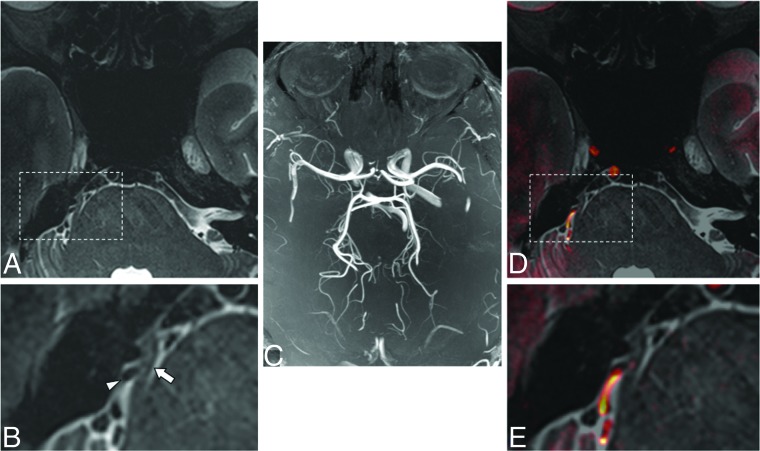
Fig 4.
Axial T2-weighted image at 7T of a patient with classic trigeminal neuralgia and associated right-sided neurovascular compression (arrow indicates right trigeminal nerve; arrowhead indicates artery in A and magnified inset B). There may be subtle hyperintensity within the nerve itself. Whole-brain maximum intensity time-of-flight projection at 7T (C) and fused gray-scale T2 and color-encoded TOF (D and E, at same locations as A and B) show orientation of the vessels and nerve and resultant neurovascular compression.

