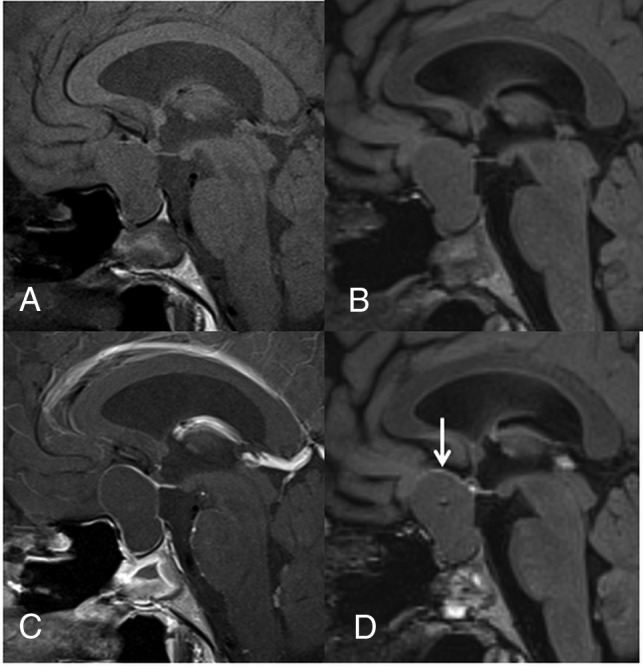
Fig 2.

A 23-year-old woman with RCC. Compared with the precontrast T1WI (A), the postcontrast T1WI (C) demonstrates enhancement in most of the cyst wall (grade 2). Compared with the precontrast 3D T2-FLAIR image (B), the postcontrast 3D T2-FLAIR image (D) shows grade 1 enhancement of some of the cyst wall (arrow). In the blinded reading study, the confidence scale of this lesion was probably an RCC (scale 2) at both the first and second interpretation sessions.
