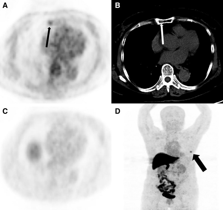
Figure 4.
A 71‐year‐old woman with newly diagnosed estrogen receptor‐positive breast cancer. (A): Axial 18F‐FDG positron emission tomography (PET) shows right manubrium focal uptake (black thin arrow). (B): Axial computed tomography (CT) show an inhomogeneous density of partial manubrium on one level (white thin arrow). (C and D): 18F‐FES PET has no uptake in the manubrium (black thick arrow shows breast mass uptake). The manubrium lesion was a considered to be a hemangioma during a recent follow‐up.

