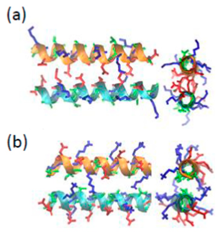
Figure 1.

The initial structures for the REMD simulations of PvLEA-22. (a) anti-parallel hydrophilic-facing model, (b) anti-parallel hydrophobic-facing model. In each figure, a side view (left) and a top view along the helix axis (right) are shown. The red, blue and green stick models represent the side chains of acidic (Asp and Glu), basic (Lys) and neutral (Ala and Thr) amino acid residues, respectively.
