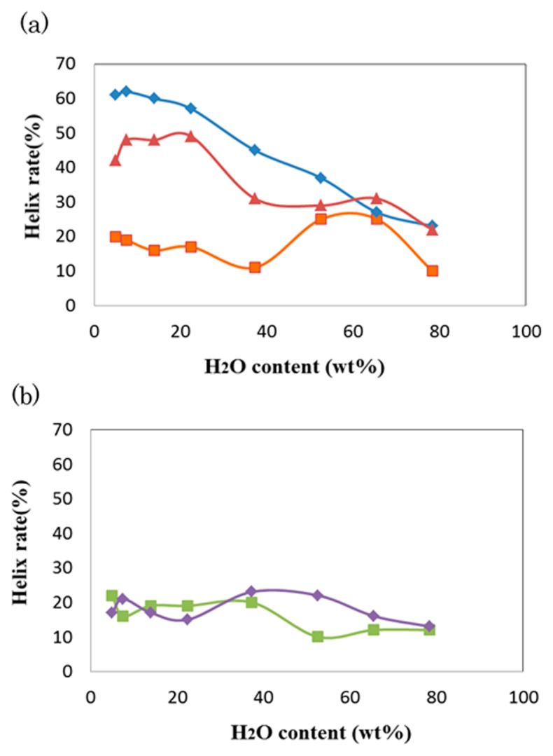
Figure 2.
Water content dependence of the helix rates of PvLEA-22. (a) The anti-parallel hydrophilic-facing (blue) and hydrophobic-facing (red) models. The results for the single-chain model are represented in orange. (b) The antiparallel double helix model (green) and single-chain model (violet) for the scrambled peptide.

