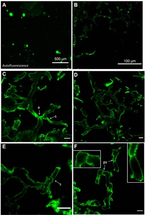
Fig. 2. Confocal laser scanning fluorescence microscopy using WGA-FITC of mycelium-like structure illustrated in Fig. 1.

Overview image in (A) shows the absence of large natural autofluorescence (i.e., without WGA-FITC) of the thin section. In contrast, with WGA-FITC labeling, the same area exhibits filamentous, mycelium-like structure visible in (B) (top section). (C to F) High-resolution confocal fluorescence views of the mycelium-like structures. The WGA-FITC binds specifically on the cell wall of the fossil filaments, which appears cylindrical (as evidenced by a rounded cross section of filaments). Several septa (s) are also stained. Note in (C) that the arrows highlight septa present in putative anastomosing filaments (a) illustrated in Fig. 1A (arrows; images are mirrored as confocal microscope is inverted). Insets in (F) illustrate septa (ps) with perforation or “bulged,” which resembles pseudosepta (39). Scale bars, 10 μm (C to F).
