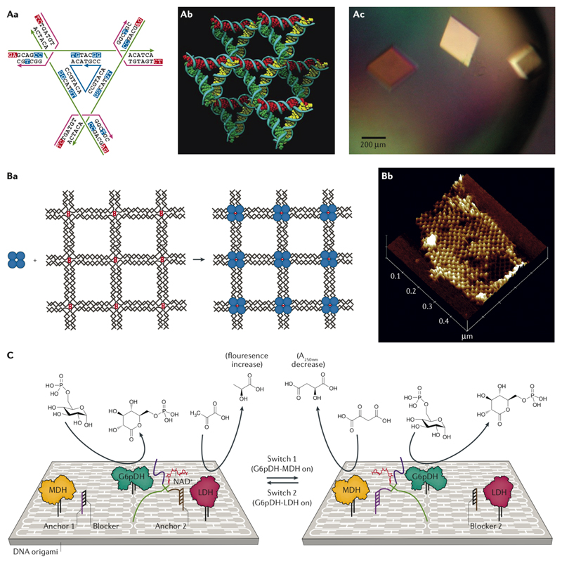
Fig. 6. Applications of DNA nanostructures.
A| The strand design of the unit cell of the tensegrity triangle crystal (Aa), the stereoscopic image of the triangles in the lattice (Ab), and the microscope image of the crystals (Ac) 100. B| Atomic force microscopy (AFM) image of streptavidin (blue circles) placed on a 2D DNA grid (Ba) and the AFM proof of assembly (Bb)35. C| Switching between two different enzyme pathways on a DNA origami support is possible by controlling the location of the cofactor (NADH)112. Panels A-C were adapted from REFs. 100, 35, 112, respectively, with permissions from Springer Nature, AAAS, and Wiley-VCH Verlag.

