
Fig. 4.
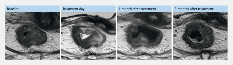
Pre- and post-MRI from patient no. 6. MR evaluation from patient no. 6 treated at Herlev hospital, Denmark. The patient had two treatments. Baseline: MRI shows a circumscript rectum tumor, thickest part 1.5 cm. Treatment day: MRI a few hours after first treatment. Only the right lateral wall was treated. The MRI shows edema in treated area. 1 month after treatment: One-month follow-up after initial treatment MRI shows decrease of tumor in the treated area. Three months after treatment: Two months after initial treatment, the patient was retreated to cover tumor in the left lateral wall. The MRI is 1 month after the second treatment and 3 months after the first treatment. The scan shows a decrease in the newly treated area but a progression in the right lateral wall.