
Fig. 5 .
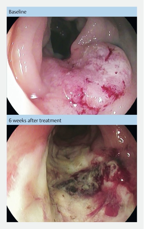
Pictures from endoscopic examination before and after treatment. Endoscopic pictures from patient no. 7 treated at Herlev hospital, Denmark. Baseline picture shows a rectal tumor estimated 3 × 2 cm. Six weeks after treatment there is a necrotic tumor decreased in size and surrounded by fibrotic tissue.