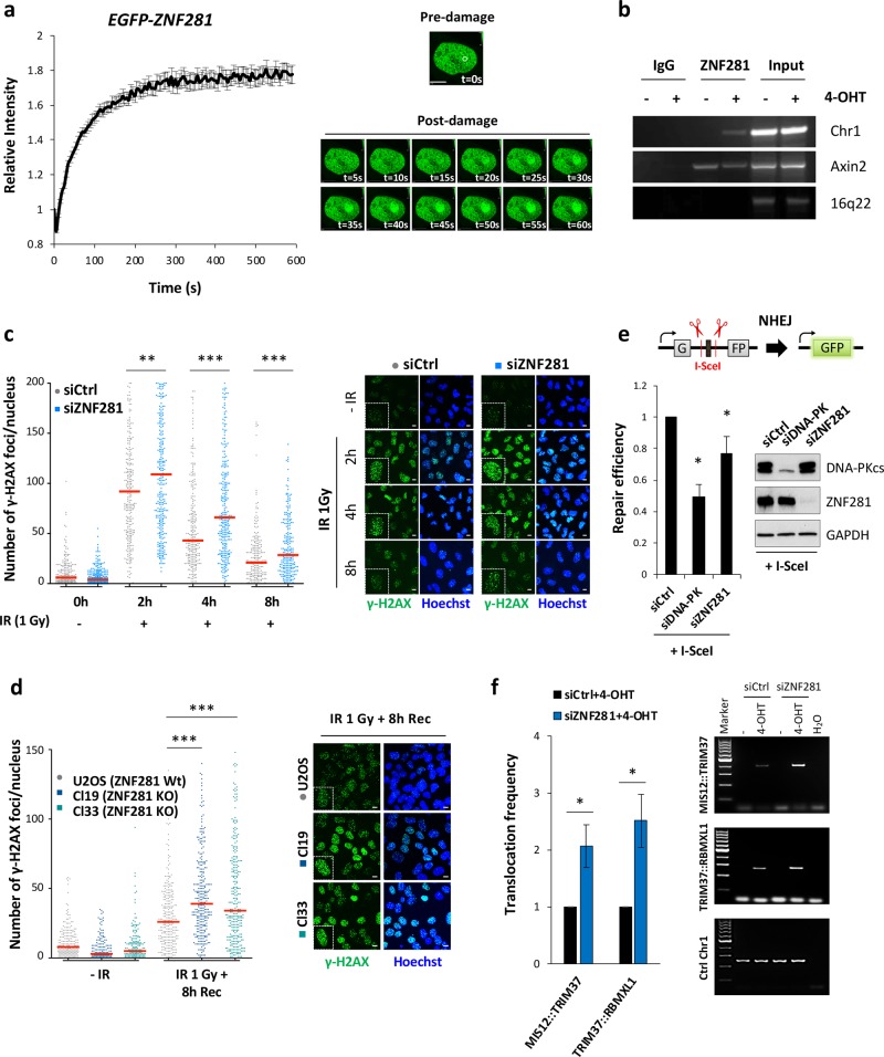
Fig. 1.
ZNF281 depletion impairs the DNA damage response and suppresses NHEJ. a EGFP–ZNF281 recruitment kinetics at DNA damage sites induced by a 355 nm laser in U2OS cells. Data were obtained from 75 cells from three independent experiments. Graphs present means ± SEM. Representative time-lapse images of the first 60 s are shown on the right. The white circle indicates the irradiated area. Scale bars, 10 μm. b ChIP analysis in U2OS-I-PpoI cells indicating the recruitment of ZNF281 to site-specific DNA break on chromosome 1 generated by I-PpoI upon activation with 1 μM 4-OHT for 2 h. Axin2 and 16q22 were used as positive and negative controls for ZNF281 ChIP, respectively; n = 2. c Number of γ-H2AX foci in U2OS cells, in which ZNF281 expression was depleted after exposure to 1 Gy of IR. After treatment, cells were allowed to recover for the indicated times and then fixed for immunofluorescence staining. Images of 200–300 cells were acquired at each time point from three biological replicates. The number of γ-H2AX foci per nucleus was automatically counted and plotted as individual data points (left panel); red lines represent medians; **p < 0.01, ***p < 0.001 (Mann–Whitney non-parametric test). Representative images are shown in the right panel. Scale bars, 10 μm. d Immunofluorescence staining for γ-H2AX in U2OS cells and in two ZNF281 knockout clones. Cells were exposed to 1 Gy of IR and then fixed after 8 h of recovery. Data obtained from at least 200 cells are plotted as individual points representing the number of γ-H2AX foci per nucleus (left panel). Red lines represent medians; n = 3; ***p < 0.001 (Mann–Whitney non-parametric test). Representative immunofluorescence images are shown (right panel). Scale bars, 10 μm. e Schematic of NHEJ reporter system (top). The presence of an adenoviral exon disrupts the GFP ORF, thus inactivating the gene. Cleavage by I-SceI and subsequent repair through the NHEJ pathway reconstitute functional GFP that is measured using FACS. Scrambled siRNA-transfected U2OS harbouring integrated NHEJ reporter cassette were used as control to measure the relative repair efficiency of DNA-PKcs- and ZNF281- depleted cells (left). Graphs present means ± SD; n = 3; *p < 0.05 (two-tailed Student’s t-test). Western blot showing the knockdown efficiency of DNA-PKcs and ZNF281 (right); GAPDH was used as a loading control. f Translocation assay in control or ZNF281-depleted DIvA cells. Upon AsiSI induction with 4-OHT, MIS12::TRIM37 and TRIM37::RBMXL1 rejoining frequencies were detected by qPCR (left panel) or end-point PCR (right panel). Amplification of an AsiSI-free region was used as a DNA input control. Graphs present means ± SEM; n = 5; *p < 0.05 (two-tailed Student’s t-test)

