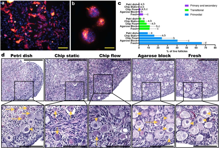
Figure 3.
Cell and ex vivo tissue culture in the Organ-on-a-chip platforms. RFP-labelled Hela cells were cultured for 4 days under perfusion in the cell culture device, displaying (a) a confluent monolayer and normal morphology in the culture chamber, and (b) spheroids/spherical aggregates in the inlet and outlet microchannels (red RFP; blue – nuclei stained with HOECHST3342). Ovarian cortical tissues from four 9- and 10-week old domestic cats were cultured for 4 days in the tissue culture device: (c) percentages of live primordial, transitional, and primary and secondary stage follicles from each treatment group, of which representative images are displayed for (d) tissue samples cultured submerged in a petri dish, in the microfluidic devices under static and flow conditions, on agarose block, and freshly collected tissues. Top scale bars (yellow) represent 200 µm and bottom ones (black) 100 µm; yellow arrowheads indicate morphologically normal, live primordial follicles, and entire arrows atretic follicle.

