Fig. 2.
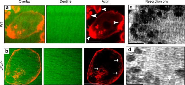
Analysis of the formation of nascent sealing zones (NSZs) and dentine resorption activity in WT and LPL−/− osteoclasts. Osteoclasts from WT (a) and LPL−/− (b) mice were cultured on dentine slices for 3 h–4 h in the presence of TNF-α and stained for actin with rhodamine-phalloidin. Confocal microscopy analysis was done in actin-stained osteoclasts. Dentine is shown in green color (pseudocolor) and actin in red. Overlay image shows the distribution of actin (red) on a dentine slice (green). Arrowheads point to NSZs in WT osteoclasts and arrows point to podosomes in LPL−/− osteoclasts. Scale bar—50 µm. Experiments were repeated three times in osteoclasts isolated from WT and LPL−/− mice. The number of NSZs were counted in ~60 WT osteoclasts total from three different experiments and the average number of NSZs is ~146 ± 21 (mean ± SD). c, d Analysis of the resorption activity in osteoclasts plated on dentine slices. Osteoclasts were cultured on dentine slices for 10 h–12 h in the presence of TNF-α. Resorption pits were scanned using a Bio-Rad confocal microscopy. Resorbed area is seen as dark areas. Arrowheads in d point to superficial pits. Scale bar—25 µm.

