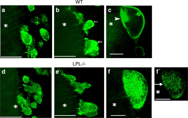
Fig. 3.
Intermittent or short time-lapse video analyses in osteoclasts from wildtype (WT) and L-plastin knockout (LPL−/−) mice. Short time-lapse video microscopy analyses at 45–60 min (a, d) 2 h–3 h (b, e), and 3 h–4 h (c, f, f′) in WT (a–c) and LPL−/− osteoclasts (d–f) expressing GFP-actin are shown. Osteoclasts were incubated with dentine slices and TNF-α during these analyses. Basolateral membrane-like structures are indicated by wavy arrows in b. NSZ is indicated by an arrowhead in c. Podosome-like structures are indicated by an arrow in f′. The asterisk in a–f′ indicates dentine matrix which is shown in diffused green color (pseudocolor). Scale bars—100 μm (a, b, d, e); 50 μm (c, f, f′).

