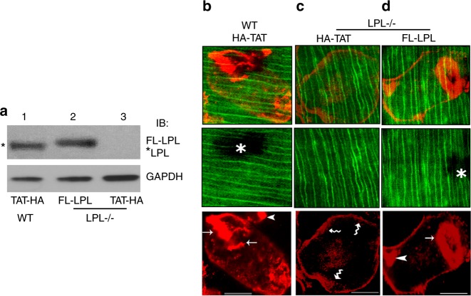
Fig. 4.
Analysis of the effect of transduction of indicated TAT-fused peptides on the formation of sealing rings in osteoclasts from wildtype (WT) and L-plastin knockout (LPL−/−) mice. a Immunoblotting analysis with an antibodyto LPL. WT and LPL−/− osteoclasts treated with bone particles and TNF-α were transduced with indicated TAT-fused FL-LPL (lane 2) or TAT-HA (lanes 1 and 3) peptide. Immunoblotting analysis with an antibody to LPL demonstrated endogenous LPL protein in WT osteoclasts (~68–70 kDa; indicated by asterisks) and transduced FL-LPL peptide in LPL−/− osteoclasts (~75–78 kDa; lane 2). Immunoblotting with a GAPDH antibody was used as loading control. b–i Osteoclasts transduced with indicated TAT-fused peptides were plated on dentine slices for 14 h–16 h in the presence of TNF-α to determine the formation of sealing rings (b–d). Confocal images of osteoclasts are shown. Merged (red and green) images are shown in the top panels. Dentine is shown in green color (pseudocolor; middle panels). An asterisk in b and d points to resorbed area underneath the osteoclast. Actin-stained cells are shown in the bottom panels. Arrows point to sealing rings and arrowheads point to NSZs. Wavy arrows point to podosomes. Scale bar—50 μm. Experiments were repeated three times with three different osteoclast preparations.

