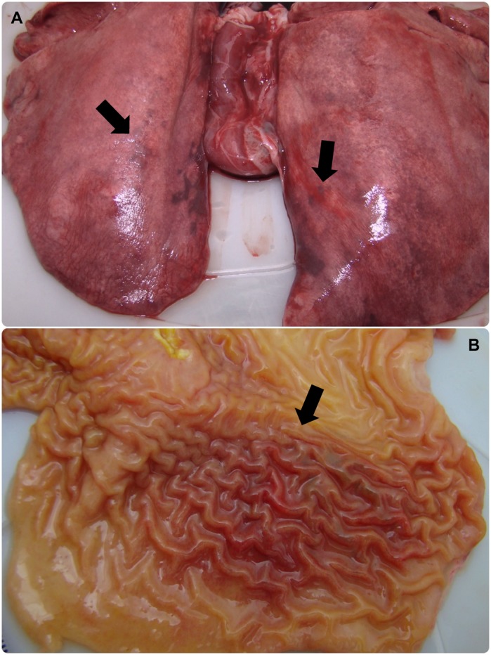
Figure 4.

Lung and stomach of capybara no. 2 after infection (phase II) with Rickettsia rickettsii (strain Itu) via tick exposure. (A) Lung of capybara no. 2 with evidence of bilateral disseminated vascular injuries (18 DPI). (B) Stomach of capybara no. 2 with an extended area of hemorrhage in the mucosa (18 DPI). This figure has been published within the Doctoral Thesis of the first author (A. Ramírez-Hernández), which is available at the University of São Paulo’s digital library of Theses and Dissertations: https://teses.usp.br/teses/disponiveis/10/10134/tde-09092019-112817/en.php.
