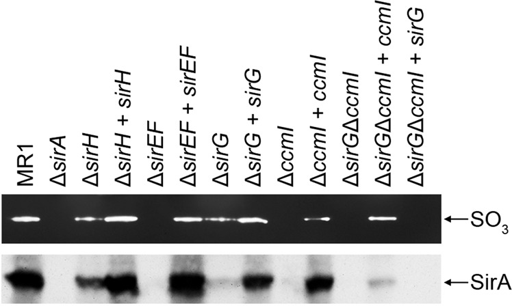
Figure 5.

Sulfite reductase activity and protein levels of wild-type, mutant, and complemented mutant cell extracts. Upper panel. Sulfite reductase activity was indicated by bands of clearing. No activity was observed in extracts from ΔsirA, ΔsirEF, ΔccmI or ΔccmIΔsirG. Reduced activity was observed in cell extracts from ΔsirH and ΔsirG. Complementation restored reductase activity to wildtype levels, except with ΔccmI, which partially restored activity. Lower panel. Western blot analysis of the cell extracts was performed using antibodies against SirA. Reactive bands that corresponded to SirA are indicated. Reduced SirA was detected in extracts from ΔsirH and ΔsirG compared to wildtype and was absent in extracts from ΔsirA, ΔsirEF, ΔccmI and ΔccmIΔsirG. Complementation restored SirA levels similar to that of the wildtype or corresponding single mutants, in the case of the ΔsirGΔccmI mutant. Full-length blots/gels are presented in Supplementary Fig. S5.
