
Supplementary Fig. 2.
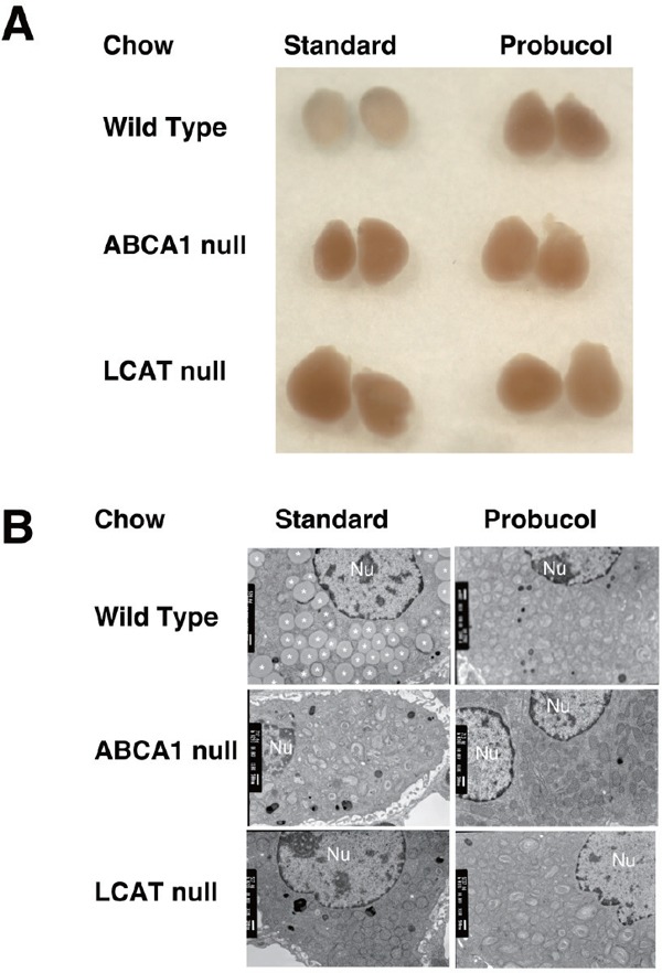
Adrenal glands of the low-HDL model mice
A. Outlook of the isolated organs. Adrenal glands were isolated from wild type, Abca1t−/−, and Lcat−/− mice, after feeding standard chow-fed or 0.5% probucol chow for 2 weeks. B. Transmitting electron microscopy of the adrenal cortex cells, from the animals fed standard chow or probucol-containing chow for 2 weeks. Nu, indicate nucleolus.