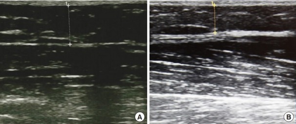
Fig. 5. Representative ultrasound images of the upper arm.

Ultrasound images of a 29-year-old woman who underwent cryolipolysis of the upper arm. A reduction in the subcutaneous fat layer under the skin was seen. (A) An ultrasound image taken prior to treatment. (B) An ultrasound image taken at the 3-month follow-up visit.
