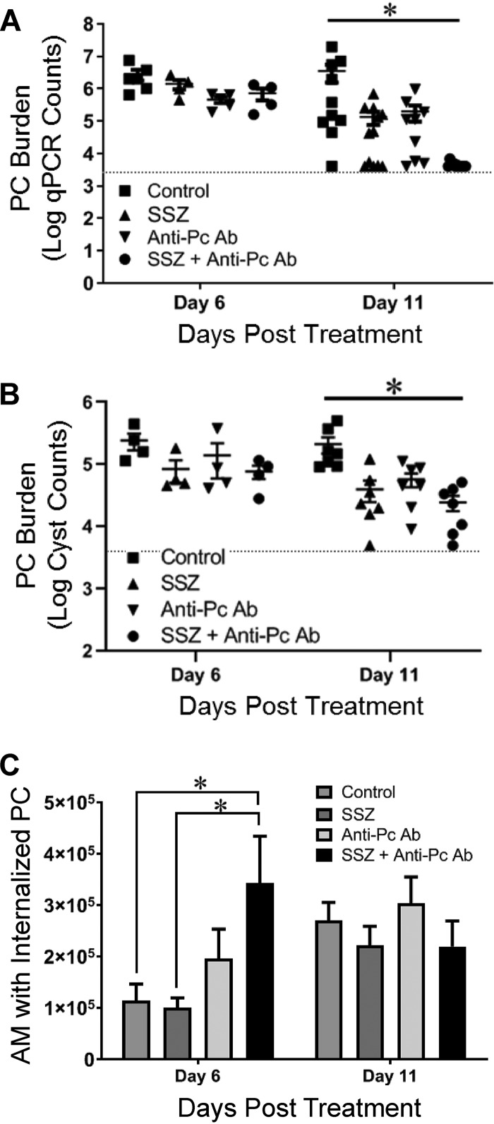
FIG 2.
Accelerated alveolar macrophage (AM)-mediated fungal clearance in anti-Pneumocystis antibody- and SSZ-treated mice. (A and B) The lung Pneumocystis (PC) burden in experimental mice was measured at 6 and 11 days posttreatment by two distinct methods: quantitative real-time PCR (qPCR) of the single-copy Pneumocystis kexin gene in lung homogenates (A) and direct Gomori’s methenamine silver staining of cysts in lung homogenate preparations (B). The limit of detection for each assay is indicated by a dotted line. Values are the mean ± 1 SEM. *, P < 0.05 between the control group and the SSZ-plus-anti-Pneumocystis antibody-treated group. At the day 6 time point, data are for 4 mice per group. At the day 11 time point, data are for ≥10 mice per group for qPCR analyses and 7 mice per group for cyst counts. (C) Macrophage phagocytosis of Pneumocystis was quantified by multispectral imaging flow cytometry, as we have described previously (15). Values are the mean ± 1 SEM. *, P < 0.05 between the indicated groups. At the day 6 time point, data are for 4 mice per group. At the day 11 time point, data are for 5 to 7 per group.

