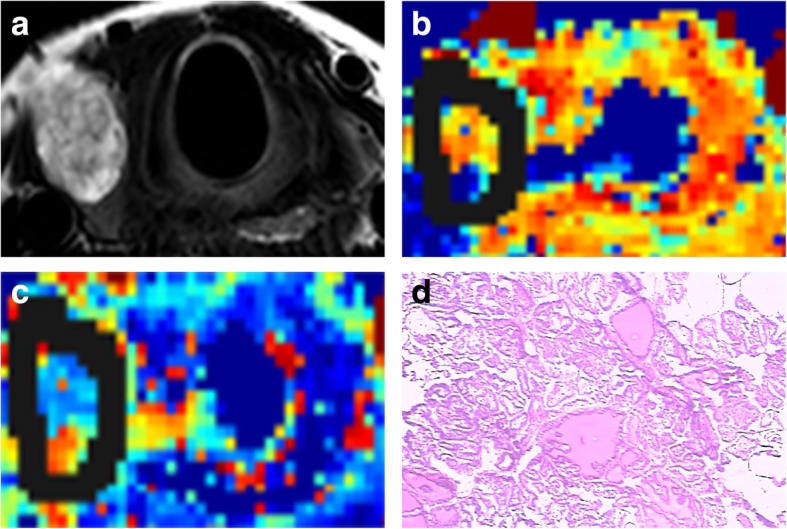
Fig. 2.

Images of a 65-year-old woman with right lobe papillary thyroid cancer (eccentric part containing cystic necrosis). a. Axial T2-weighted image. b-c. IVIM colour map (D and f). D = 0.83 × 10− 3 mm2/s, f = 32.15%. Solid lines show borders containing cystic necrosis. d. Nuclei arranged closely in papillary thyroid carcinoma (HE, × 100)
