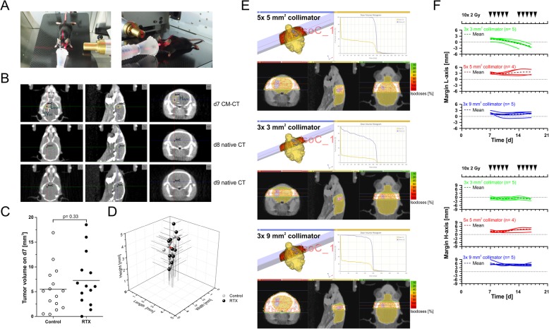
Fig. 2.
Contrast-enhanced and native CBCT scans for tumor localization, tumor volume follow-up, treatment planning, dose administration, and repositioning of animals. a Positioning and fixation of the mouse inside an anaesthetic mask with an elastic membrane. b Alternating contrast-enhanced (d7) and native CBCT scans (d8 and d9) for tumor localization, tumor volume follow-up, treatment planning, dose administration, and repositioning of animals. The black cross marks the isocenter defined as the center of the contrast-enriching volume on d7 and inferred from its relative position to bony structures in native CT scans on d8/d9. c Tumor volumes of irradiated and non-irradiated animals at the start of treatment (d7) as determined by Lx Hx W calculations shown in (b). p-value as calculated by exact Wilcoxon Rank test. d Tumor measures (L, H, and W) of individual animals at the start of treatment (d7). The red arrowhead indicates the animal shown in (b) and (e). e Treatment plans and dose-volume histograms for irradiation with two transversal, contralateral beams of 5 × 5, 3 × 3, or 3× 9 mm2 collimation, respectively. f Analyses of margins between contrast enriching tumor volumes and beam collimation settings for all irradiated animals in L- and H-axis over time

