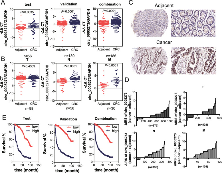
Fig. 2.
CircPTK2 is overexpressed in tissues of CRC patients and associated with tumor metastasis. a Levels of circPTK2 in the indicated fresh tissues from the testing (n = 56), validation (n = 130), and the combination set (n = 186) were detected by qPCR. The circPTK2 expression was normalized to GAPDH. b The abundance of circPTK2 in CRC patients with different clinical characteristics were further analyzed. T: primary tumor; N: node metastasis; M: distant metastasis. Data are presented as -ΔΔCt by one-way ANOVA with three technical replicates each. c The levels of circPTK2 in TMA were evaluated by RNA-ISH analysis, and the representative staining images are shown. d The difference in staining score between CRC lesions and adjacent non-tumor tissues in total (n = 973), tumor (T) (n = 528), node (N) (n = 285) and metastasis (M) (n = 110) cohort are shown. e Kaplan–Meier curves depicts the overall survival of CRC patients according to the tumoral circPTK2 levels. Left to right: testing cohort, validation cohort and combination cohort. P values were calculated by the log-rank test

