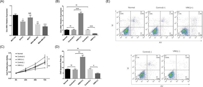
Figure 4.

Circ‐VRK1 in cell experiments. Circ‐VRK1 expression was reduced in BT474, MDA‐MB‐453 and MDA‐MB‐231 cell lines but unchanged in MCF7 cell line compared with normal breast epithelial cell line MCF10A (A). Circ‐VRK1 expression was upregulated in the VRK1 (+) group and downregulated in the VRK1 (−) group (B). Cell proliferation was reduced in the VRK (+) group but increased in the VRK (−) group (C). Cell apoptosis rate was increased in the VRK1 (+) group but decreased in the VRK1 (−) group (D, E). Comparison of circ‐VRK1 expression between the normal cell line and each breast cancer cell line was carried out by t test. Comparison of circ‐VRK1 expression, cell proliferation ability and cell apoptosis between the VRK1(+) group and control (+) group as well as the VRK1(−) group and control (−) group was determined by t test. P < .05 was considered significant. *P < .05 and **P < .01. VRK(+), circ‐VRK1 overexpression; control (+), blank overexpression; control (−), blank shRNA; VRK (−), circ‐VRK1 shRNA; circ‐VRK1, circular RNA VRK serine/threonine kinase 1
