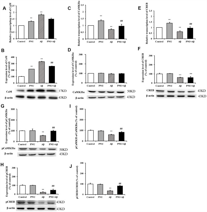
Figure 10.
Activation of α7 nAChR activates the CaM-CaMKII-CREB signaling pathway in Aβ oligomer-treated neurons. The x-axis labels are Control, hippocampus cells from the WT rat; PNU, WT hippocampus cells treated with PNU; Aβ, the WT hippocampus cells treated with Aβ; and PNU+Aβ, the WT hippocampus cells treated with PNU and Aβ. The y-axis indicates the relative mRNA or protein levels as a percentage of the control. mRNA and protein expression levels in each group were measured by RT-qPCR and western blot analysis, respectively. Detection of the levels of CaM (A) mRNA and (B) protein, CaMKIIα (C) mRNA and (D) protein, CREB (E) mRNA and (F) protein, (G) p-CaMKIIα protein and (H) p-CREB protein, and the (I) p-CaMKIIα/CaMKIIα and (J) p-CREB/CREB ratios. The results demonstrated that the transcription of CaM and the protein level of CaM were significantly increased in the Aβ group, while the expression level of α7 nAChR was decreased. The transcription level of CaMKIIα was significantly decreased, and the expression levels of p-CaMKIIα, CREB and p-CREB, and p-CaMKIIα/CaMKIIα and p-CREB/CREB ratios were significantly decreased in the Aβ group. All these protein levels were largely restored following activation of α7 nAChR by PNU treatment. Data are presented as the mean ± standard deviation. *P<0.05, **P<0.01 vs. Control group; #P<0.05, ##P<0.01 vs. Aβ.

