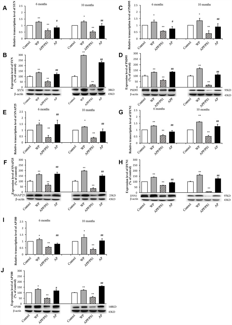
Figure 5.
Activation of α7 nAChR increases the expression of synaptic-associated proteins in the hippocampus of APP/PS1_DT mice. The x-axes are the WT mice (control), the WT mice treated with PNU (WP), the APP/PS1_DT mice (APP/PS1) and the APP/PS1_DT mice treated with PNU (AP). The y-axes are the relative level of mRNA or protein (% of control group). Detection of SYN (A) mRNA and (B) protein; PSD95 (C) mRNA and (D) protein; SNAP25 (E) mRNA and (F) protein; DYN1 (G) mRNA and (H) protein; AP180 (I) mRNA and (J) protein by RT-qPCR and western blot analysis. Protein expression levels were detected by western blot analysis (β-actin was used as an internal control). RT-qPCR and western blot analysis demonstrated that the expression levels of SYN, PSD95, SNAP25, DYN1 and AP180 in the hippocampus of APP/PS1_DT mice were significantly decreased compared with the control group, and this decreasing trend was partially reversed by PNU treatment. Data are presented as the mean ± standard deviation. *P<0.05, **P<0.01 vs. control group; #P<0.05, ##P<0.01 vs. APP/PS1 group.

