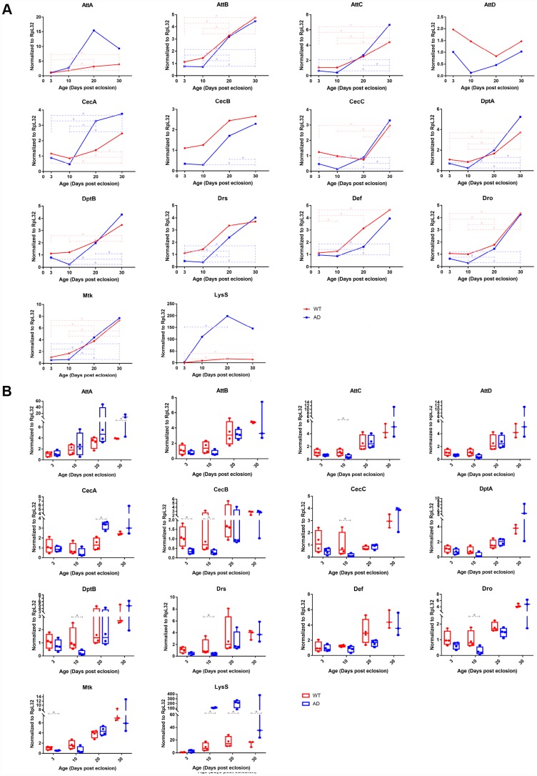
Figure 3.
Quantitative PCR validation of differentially expressed immune-related genes in control and Aβ transgenic flies. The results confirmed age-associated alterations in expression trends and transcriptional regulatory levels among the AMP and LysS genes between healthy control and disease model flies. The line chart (A) displays the time series (3-, 10-, 20-, and 30-days post eclosion) gene expression in the head tissue of WT (red, round dots) and AD (blue, square dots) flies. The box plot (B) exhibits the comparison of mRNA levels between normal (red, left) and disease (blue, right) model flies among the age groups.

