Figure 1.
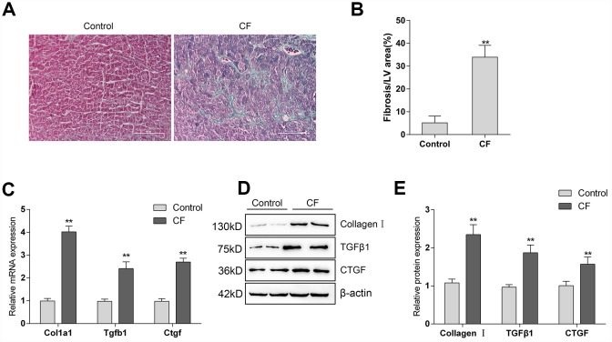
The construction and identification of ISP-induced cardiac fibrosis (CF) model in rats (A) Pathomorphological features of rat hearts in different groups examined by Masson staining. (B) In Masson staining, fibrotic tissue was stained to blue and myocardium to red. The fibrotic areas were calculated and the percentage of fibrotic tissue area was used to assess CF. (C–E) The mRNA expression and protein levels of Col1a1 (Collagen I), Tgfb1 (TGFβ1), and Ctgf (CTGF) in CF and control groups determined by real-time PCR and Immunoblotting analyses.

