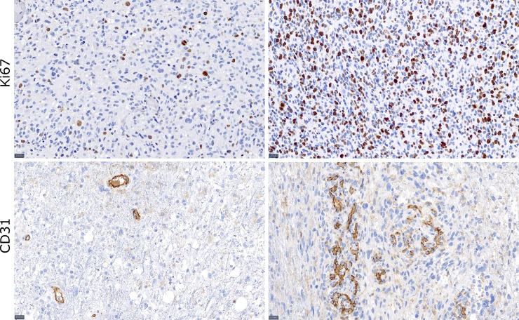
Fig 3. Exemplary histologic specimens of targeted tumor biopsies in a 20-fold magnification (black bars equal 20 μm).
The upper row shows Ki67 immunohistochemistry to determine the proliferative activity with a low percentage of 8% proliferating cells (left) and high percentage of 40% proliferating cells (right). The lower row shows CD31 immunohistochemistry to determine the amount of endothelial cells with a low vessel density (score 1, left) and a high vessel density (score 3, right).

