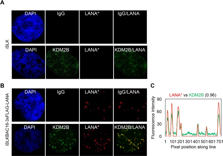
Fig 5. Co-localization of KDM2B with LANA in latent KSHV-infected cells.
(A) Uninfected iSLK cells or (B) KSHV-infected iSLK cells (iSLKBAC16-3xFLAG-LANA) were subjected to immunofluorescence analysis for LANA (red) and KDM2B (green). Rabbit IgG served as a negative control. FLAG antibody was used to detect 3xFLAG-LANA expressed from KSHV. (C) Representative LANA puncta were connected by yellow line and the co-localization of LANA with KDM2B along the line was analyzed by ImageJ.

