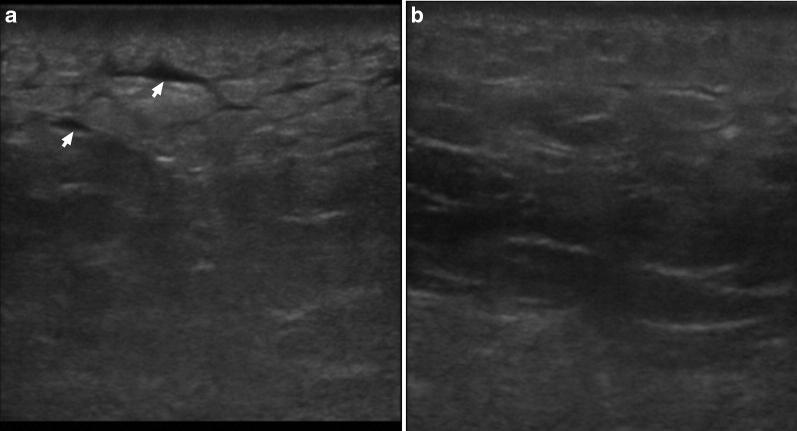
Fig. 4.
Point-of-care ultrasound scan of the affected left gluteal soft tissue 4 days after admission (a) and 8 days after admission (b) using a 15–6 MHz linear probe. These images show the progression of the sonographic appearance of NSTI, initially with accumulation of fluid in the subcutaneous tissue (arrows), giving it a “cobblestone” appearance and a progressive return to the normal architecture of the subcutaneous tissue over time

