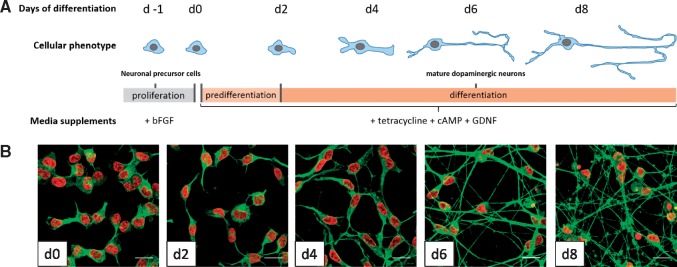
Figure 1.
Differentiation of LUHMES cells. (A) Schematic representation of the differentiation procedure showing phenotypic changes during the differentiation from neuronal precursor cells to mature dopaminergic neurons. (B) Representative fluorescent confocal microscopy images of LUHMES immunostained during different stages of maturation (d0–d8 of differentiation) for βIII-tubulin (green). Nuclei are labeled by DNA staining with H-33341 dye (red). Scale bar = 20 µm.

