Figure 1.

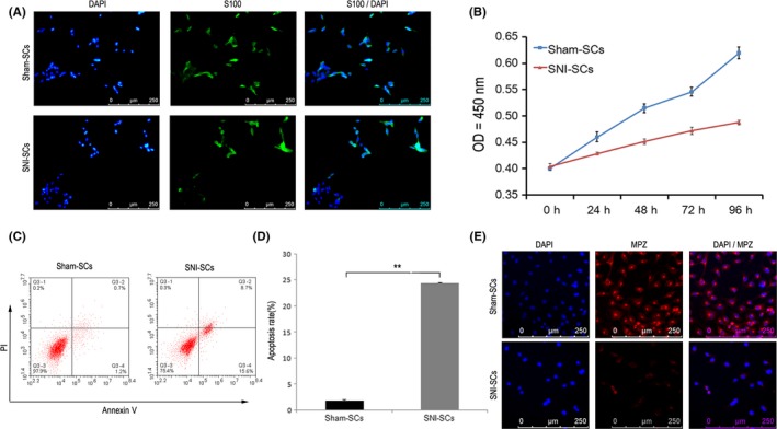
Decrease in SC proliferation and increase in SC apoptosis following sciatic nerve injury. A, Immunofluorescence staining showed that Sham‐SCs and SNI‐SCs express the SC marker S100. Scale bar: 20 µm. B, The cell proliferation curve of Sham‐SCs and SNI‐SCs, according to the CCK8 assay findings. Repeated measurement variance analysis, F = 254.067, P < 0.001. C, Apoptosis of Sham‐SCs and SNI‐SCs, as demonstrated by Annexin V and PI staining analysis. D, The apoptosis rate of SNI‐SCs was significantly increased as compared to Sham‐SCs. Data represent the mean ± SD values. **P < 0.01, Student's t test. E, Immunofluorescence staining showed that the expression of MPZ was lower in SNI‐SCs than in Sham‐SCs. Scale bar: 20 µm
