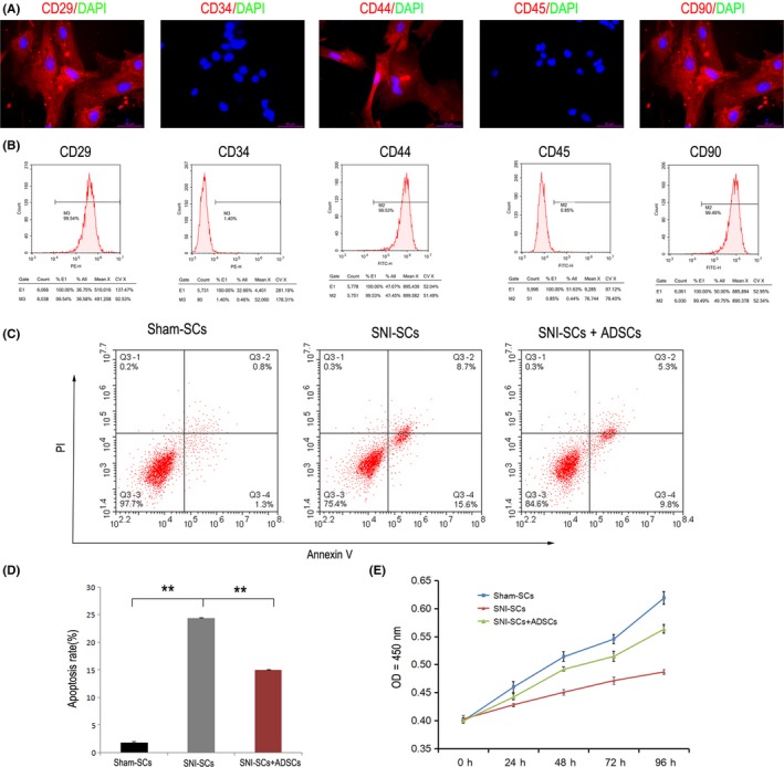
Figure 2.

Decrease in SC apoptosis and increase in SC proliferation after coculture with ADSCs. A, Immunofluorescence staining showed that ADSCs expressed CD29, CD44, and CD90, but they did not express CD34 and CD45. Scale bar: 20 µm. B, Flow cytometry analysis of apoptosis. C, Apoptosis of Sham‐SCs, SNI‐SCs, and SNI‐SCs + ADSCs was analyzed by Annexin V and PI staining. D, The apoptosis rate of SNI‐SCs decreased significantly after treatment with ADSCs. Data represent the mean ± SD values. **P < 0.01, one‐way ANOVA. E, The cell proliferation curve of Sham‐SCs, SNI‐SCs, and SNI‐SCs + ADSCs, as measured by the CCK8 assay. Repeated measurement variance analysis, F = 204.983, P < 0.001. LSD t test, P < 0.05
