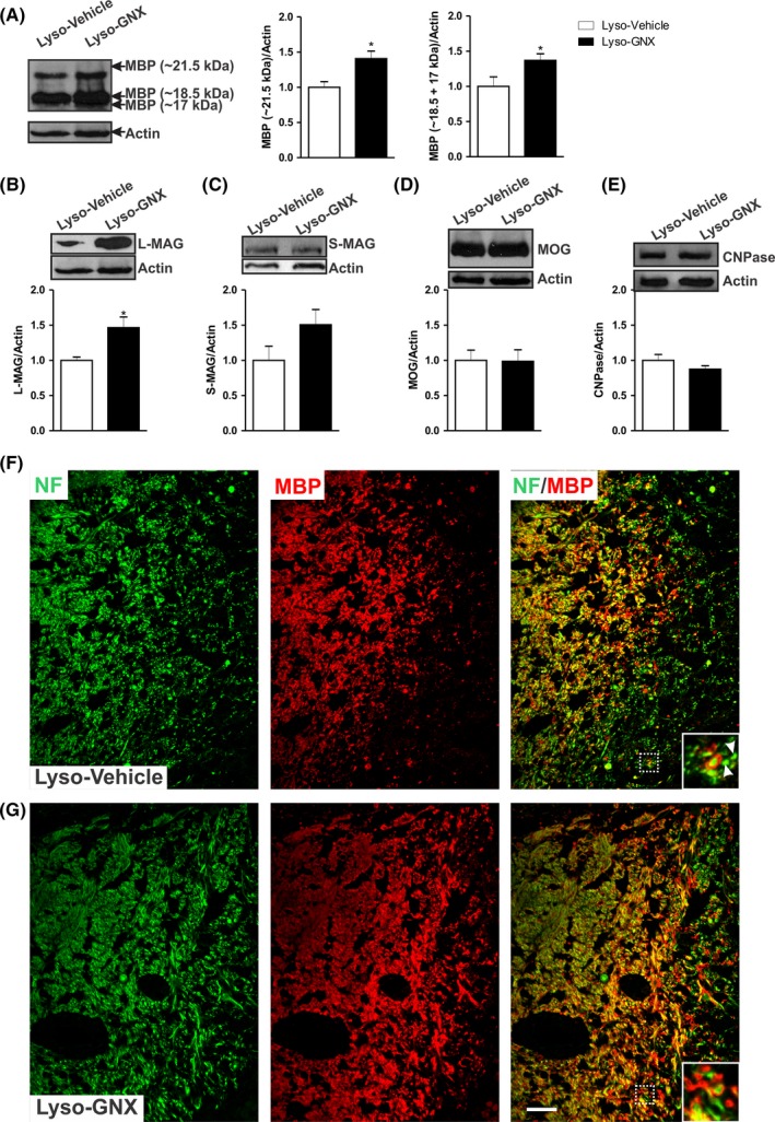
Figure 3.

GNX promotes the expression of major myelin proteins 7 days post–lysolecithin‐induced demyelination in the corpus callosum. A, GNX treatment significantly increased the expression levels of the 21.5 kDa and the 18.5 + 17 kDa isoforms (Lyso‐Vehicle: n = 5, Lyso‐GNX: n = 4, P < .05). B, There was a significant increase in the expression of L‐MAG in GNX‐treated rats compared to vehicle‐treated rats (P < .05). Expression levels of S‐MAG (C), MOG (D), and CNPase (E) were not affected by GNX. (F) and (G) are immunofluorescent staining images of NF (green) and MBP (red) in the corpus callosum of vehicle‐treated and GNX‐treated OVX rats, respectively, 7 days postdemyelination. Inserts show higher magnifications of the area delimited by the white box. The axons (green) are surrounded by the myelin sheath (red). The arrowheads in (F) point to axons devoid of myelin. GNX treatment shows better axonal remyelination compared to vehicle‐treated rats. Scale bar = 50 µm
