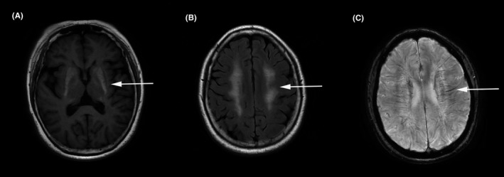
Figure 1.

Brain magnetic resonance imaging showed lesions in the bilateral basal ganglia with short T1 signals (A, arrow), lesions across the bilateral centrum semiovale with long T2 signals (B, arrow), as well as slight dilations of the deep medullary vein along the bilateral periventricles on susceptibility weighted imaging (C, arrow)
