Figure 2.

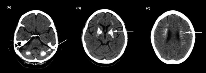
Head computed tomography scan showed symmetric calcified lesions in the dentate nucleus (A, arrow) and lenticular nucleus (B, arrow) bilaterally, as well as radiating high‐density lesions along the periventricular region (C, arrow)

Head computed tomography scan showed symmetric calcified lesions in the dentate nucleus (A, arrow) and lenticular nucleus (B, arrow) bilaterally, as well as radiating high‐density lesions along the periventricular region (C, arrow)