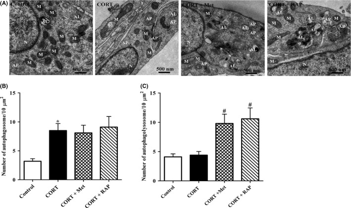
Figure 5.

Effects of treatment with Met or RAP on the ultrastructures of PC12 cells induced by high CORT. A, Representative TEM images of PC12 cells. PC12 cells were incubated with CORT (10 μmol/L) in the presence of Met (2 mmol/L) or RAP (200 nmol/L) for 24 h. Cells were then harvested for TEM observation as described in Section 2. Mitochondria (M), nucleus (N), autophagosome (AP), and autolysosomes (ALs) were indicated. B, C, The number of APs and ALs was quantified (n = 12 cells/group). All the data were presented as mean ± SEM. *P < .05 vs control group; # P < .05 vs CORT group. Scale bar, 500 nm
