
Figure 4.
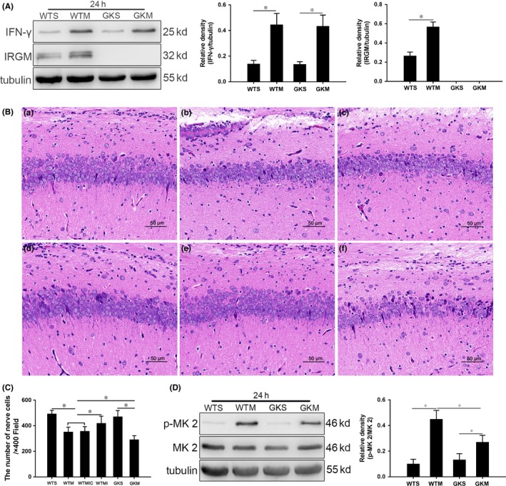
Pathological changes in the hippocampus of mice with SAE are aggravated, and p38 MAPK signaling is inhibited in IRGM1 knockout mice. A, Expression of IFN‐γ and IRGM1 in the hippocampus at 24 h quantitated by Western blotting. Protein levels are normalized to those of β‐tubulin and are shown as relative arbitrary units. B, Pathological changes in the hippocampus of mice 24 h postsurgery. (a) Relatively normal morphology in the WTS group; in (b) WTM and (c) WTMIC groups, some cells dissolve and numbers of cells decrease; (d) in the WTMI group, cells are irregular in shape and somewhat disordered in arrangement; (e) in the GKS group, morphology is relatively normal; and (f) in the GKM group, cells are reduced and disordered, presenting as acute traumatic changes. Scale bar = 50 μm. C, Cell counts from HE‐stained sections. D, Expression of p‐MK 2 and MK 2 at 24 h quantitated by Western blotting. Protein levels are normalized to those of β‐tubulin and shown as relative arbitrary units. Data are from 3 independent tests, n = 5 for each group, *P < .05