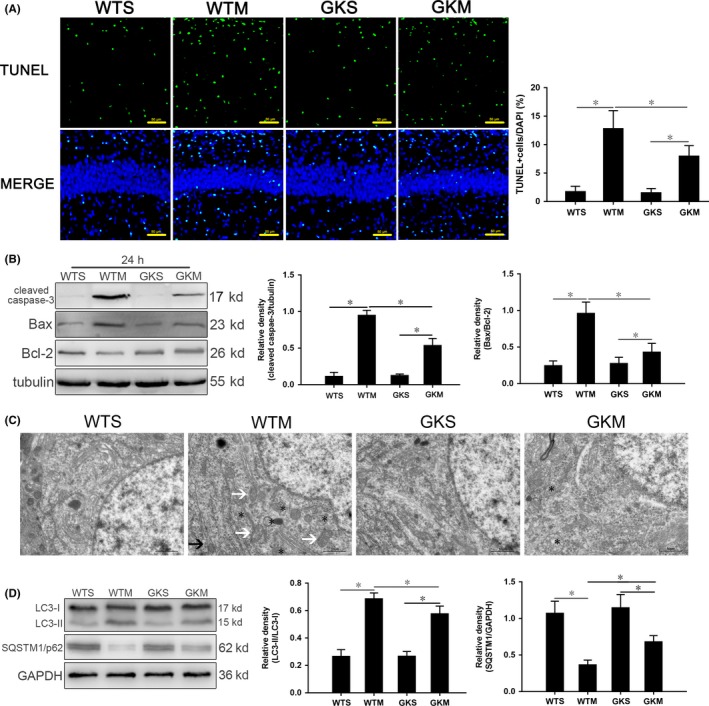
Figure 5.

The absence of IRGM1 reduced apoptosis and autophagy in mice with SAE. A, TUNEL staining; apoptotic bodies are green, and nuclei are blue. Scale bar = 50 μm. B, Expression of cleaved caspase‐3, Bcl‐2, and Bax quantitated by Western blotting. Protein levels are normalized to those of β‐tubulin and shown as relative arbitrary units. C, TEM showing lack of autophagosomes in the WTS and GKM groups; in the WTM group, there were many autophagosomes enclosing damaged organelles and proteins (*); and in the GKM group, there were a few autophagosomes (*) and no complete intracellular structures were observed. Scale bar = 10 μm. D, Expression of LC3 and SQSTM1/p62 quantitated by Western blotting. Protein levels are normalized to those of GAPDH and shown as relative arbitrary units. Data are from 3 independent tests, n = 5 for each group, *P < .05
