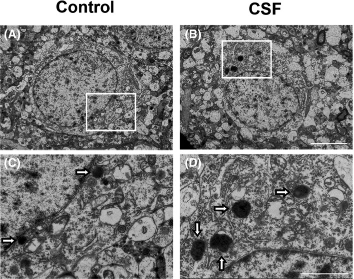
Figure 4.

CSF caused enlargement and increase in intracellular lysosomes in the cortex detected by electron microscopy. A‐B, Representative electron photomicrographs of neurons from mice in the CSF and control groups. Scale bar = 5 μm. C‐D, Higher magnification images of the rectangle area in (A‐B), hollow arrows marked intracellular lysosomes. Scale bar = 2 μm
