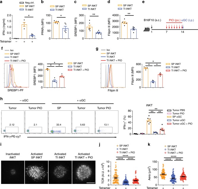
Fig. 6. PIO restores cholesterol and IFN-γ production in tumor-infiltrating iNKT cells in vivo.
a IFN-γ production in tumor-infiltrating (TI) iNKT cells and splenic (SP) iNKT cells from B16F10 tumor-bearing mice, after activating with plate-coated mCD1d-PBS57 tetramer. b–d PPARγ (b), SREBP1 (c), and Filipin III staining (d) in TI iNKT cells and SP iNKT cells from B16F10 tumor-bearing mice. e Timeline of experimental procedure. f–h SREBP1 expression (f) and Filipin III staining (g) of iNKT cells, and percentages of IFN-γ+ iNKT cells (h) from indicated tissues of B16F10 tumor-bearing mice, after treating with or without PIO for 7 days and injecting with 2 μg αGC for 5 h. i–k Distribution (i) and fluorescence intensity (j) of surface TCR, and area of immunological synapse (k) of iNKT cells, after activating with coverslip-coated CD1d-PBS57 tetramer for 45 min. Cells from indicated tissues were isolated from mice treating with or without PIO for 7 days (n = 80 cells per group). Bar, 5 μm. Data are means ± SEM of four mice (a, f, g), five mice (b, d), or six mice (c, h), pooled from three independent experiments. Data were analyzed by Mann–Whitney test (a–d, f–h) or unpaired Student’s t-test (j, k). *P < 0.05, **P < 0.01, ****P < 0.0001. Source data are provided as a Source Data file.

