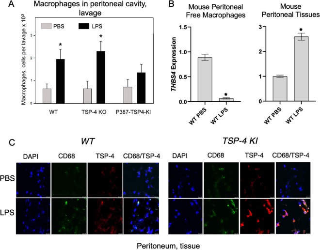
Fig. 6. TSP-4 promotes accumulation of macrophages in peritoneal tissue of mice with LPS-induced peritonitis.
a The number of macrophages in peritoneal cavity in mice with LPS-induced peritonitis. *p < 0.05, n = 5. b TSP-4 expression in macrophages from the peritoneal cavity lavage (left panel) and in the peritoneal tissue. QRT-PCR, fold increase (RQ) over the values in control mice injected with PBS; n = 3; *p < 0.05. c Macrophages and TSP-4 in peritoneal tissue of WT and P387-TSP-4-KI mice with LPS-induced peritonitis. Immunofluorescence; blue = nuclei (DAPI), green = macrophages (anti-CD68), red = TSP-4 (anti-TSP-4). Scale bar is 20 µm.

