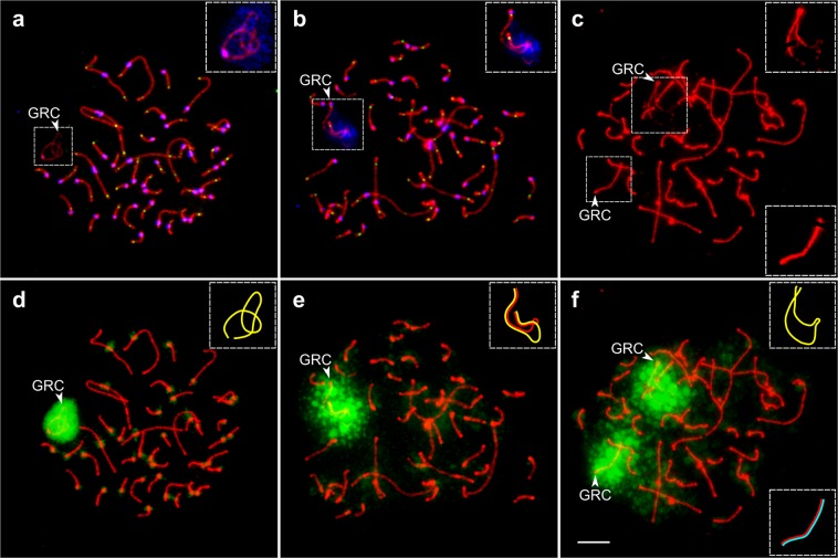
Figure 4.
Pale martin pachytene spermatocytes with one (a,d), two (b,e), and three (c,f) copies of GRC. (a–c) Cells after immunostaining with antibodies against SYCP3 (red), centromere proteins (blue) (a,b) and MLH1 (green) (a,b). (d–f) The same cells after FISH with the pale martin GRC paint probe. Arrowheads point to GRCs. Inserts show zooms at the GRC with enhanced brightness and contrast (a–c) and schematic representations of GRC SCs (d–f). Note MLH1 foci at both ends of the partially paired GRC bivalent (b). Bar – 5 µm.

