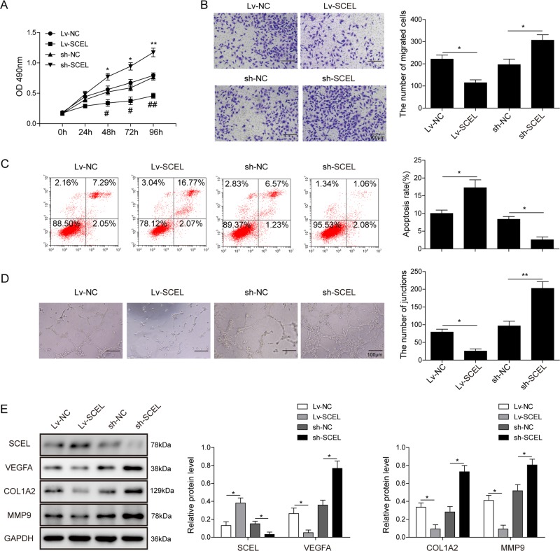
Fig. 7. SCEL inhibits cell proliferation, migration, angiogenesis, and promotes apoptosis in HUVECs.
a Cell viability was monitored by the MTT assay. b Cell migration capacity was monitored by the transwell migration assay. Data were representative images or were expressed as the mean ± SD of n = 3 experiments. c Cell apoptosis was determined by Annexin-V-FITC/PI staining followed by flow cytometry. d In vitro angiogenesis was quantified by tube formation assays. e Protein levels of Col1a2, MMP9, and VEGFA were determined by western blotting. GAPDH served as loading control. Data were representative images or were expressed as the mean ± SD of n = 3 experiments. *P < 0.05 and **P < 0.01.

