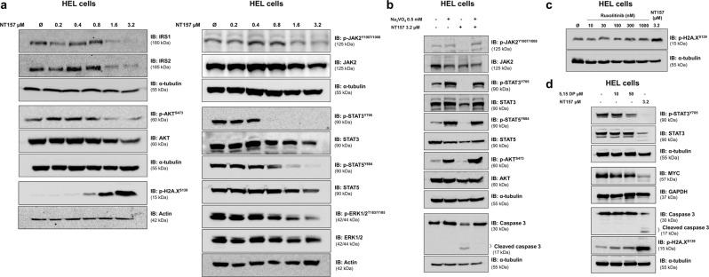
Fig. 2. NT157 downregulates IRS1/2, JAK2/STAT, AKT, and ERK1/2 signaling and induces DNA damage in HEL cells.
a Western blot analysis is shown for IRS1, IRS2, p-AKTS473, AKT, p-H2A.XS139, p-JAK2T1007/1008, JAK2, p-STAT3Y705, STAT3, p-STAT5Y694, STAT5, p-ERK1/2T183/Y185, and ERK1/2 levels in total cell extracts from HEL cells treated with NT157 (Ø, 0.2, 0.4, 0.8, 1.6, and 3.2 µM) for 24 h. b Western blot analysis is shown for p-JAK2T1007/1008, JAK2, p-STAT3Y705, STAT3, p-STAT5Y694, STAT5, p-AKTS473, AKT, and caspase 3 levels in total cell extracts from HEL cells previously treated with Na3VO4 (0.5 mM) for 30 min followed by NT157 (3.2 µM) treatment for an additional 24 h as indicated. c Western blot analysis is shown for p-H2A.XS139 from HEL cells treated with ruxolitinib (Ø, 10, 30, 100, 300, and 1000 nM) or NT157 3.2 µM for 24 h. d Western blot analysis is shown for p-STAT3Y705, STAT3, MYC, caspase 3, and p-H2A.XS139 levels in total cell extracts from HEL cells treated with 5,15-DP (Ø, 10 and 50 µM) or NT157 (3.2 µM) for 24 h. Membranes were reprobed with an antibody for the detection of total target protein and/or actin, GAPDH or α-tubulin, and images were detected with a SuperSignal™ West Dura Extended Duration Substrate system using a Gel Doc XR+ imaging system.

