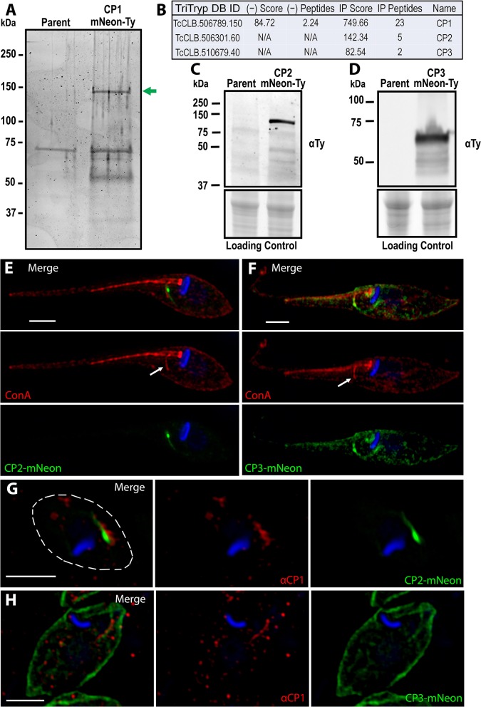
Figure 4.
Identification of the CP1 associated proteins CP2 and CP3. (A) SDS page gel of CP1-mNeon-Ty CoIP eluate stained for total protein. Several bands are present in the CoIP that are absent from the control IP using parental lysate. Band excision and MALDI mass-spectroscopy (MS) analysis confirmed that the upper band (green arrow) is CP1-mNeon-Ty. (B) Notable hits from Orbitrap shotgun MS of the CP1 CoIP eluate revealed two hypothetical protein (CP2, CP3) in addition to CP1. αTy immunoblots of lysates from CP2-mNeon-Ty (C) and CP3-mNeon-Ty (D) overexpressing epimastigotes show that the tagged protein are expressed. (E,F) SR-SIM of CP2-mNeon-Ty and CP3-mNeon-Ty overexpressing mutants showing SPC labeling. 4°C labeling of with Concanavalin A -Rhodamine labels the SPC pre-oral ridge (white arrows). CP2-mNeon-Ty (G) and CP3-mNeon-Ty (H) in amastigotes also localize to the SPC, labeled by αCP1. Scale bars: 2 μm.

