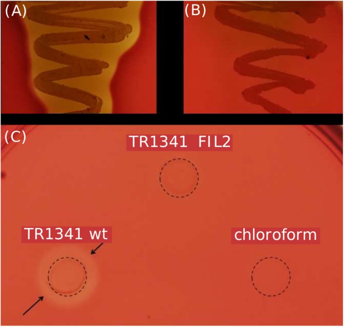
FIGURE 2.
β-hemolytic features of S. sp. TR1341. (A) β-hemolysis of TR1341 wt colonies in a standard blood agar plate, 3 days incubation in 28°C. (B) Disruption of the filipin/fungichromin production in TR1341ΔFIL2 causes complete loss of β-hemolysis. Grown for 4 days at 28°C in blood agar. (C) Production of hemolytic secondary metabolites by the strain (TR1341 wt) – extracted from 3-days old culture grown in GYM media by acetone/ethylacetate liquid phase extraction. The amount of extract applied (10 μl) corresponds to 4 ml of original culture. Dashed circle indicates the extract drop border, the arrows show the hemolytic zone. TR1341ΔFIL2 is the extract of filipin-deficient mutant of TR1341, chloroform stands for the negative control (the solvent only). Photographed after 16 h incubation at 28°C.

