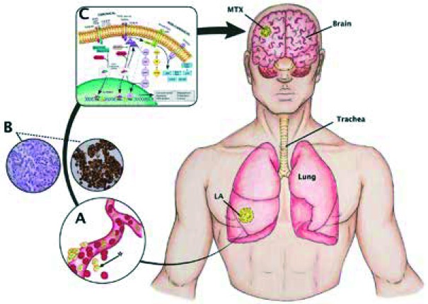
Fig. 1.
Canonical (Smad-dependent) and non-canonical (Smad-independent) pathways. An oversimplified and modified (from [30]) scheme depicting the TGF-β pathway as well as a model of cerebral metastasis from lung adenocarcinoma. A) Disaggregation of cells from primary tumour mass to a particular metastatic niche is mediated mainly through integrins and proteases expressed at the surface of cancer cells, vasculature, and stromal cells [48]. In summary, cells [*] break through the vascular basement membrane via the expression of cellular matrix metalloproteinase activity – MMP2 and MMP9. Many angiogenic factors secreted by both tumour and stromal cells, such as VEGF and platelet-derived growth factor β (PDGF-β), which are transcriptionally directly driven by hypoxia-inducible factor 1 or 2 (HIF1–HIF2). B) Lung adenocarcinoma is commonly defined as a slow-growing cancer that arises mostly from distal airways, typically with glandular histology displaying biomarkers that are consistent with an origin in the distal lung, including thyroid transcription factor 1 (NKX2-1) and keratin (7) with complex early diagnosis because it usually involves the periphery of the lung and has scarce symptoms, promoting early metastasis. C) The TGF-β1 signalling is regarded as an initial, complex, and yet crucial cytokine to induce EMT during cancer progression and metastasis (20) in a Smad2/Smad3 complex-dependent C-terminal phosphorylated downstream manner via ALK-5 receptors, and also dependent on the protein-serine/threonine kinases that participate in the Ras-Raf-MEK5-ERK5 signal transduction cascade and nuclear translocation

