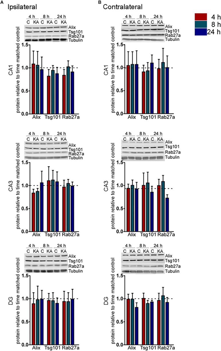
FIGURE 2.
Acute regulation of protein levels of exosome biogenesis pathways after status epilepticus in hippocampal subfields. Representative immunoblots and graphs show protein levels of Alix, Tsg101, and Rab27a exosome pathway components 4, 8, and 24 h after SE compared to control (C) in each subfield of the (A) ipsilateral and (B) contralateral, hippocampus. Graphs show mean ± SEM. Protein levels were normalized to tubulin (n = 4–5 per group; ANOVA with Bonferroni post hoc test showed no significant differences).

