Figure 4.
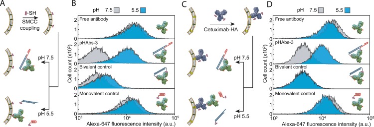
pH-dependent targeting of antibodies to human carcinoma cells. (A) Schematic representation of direct low pH triggered targeting of mammalian cells. First, A431 epidermoid carcinoma cells are covalently prelabeled with HA peptide epitopes via a Sulfo-SMCC cross-linker. The prelabeled cells are subsequently resuspended in PBS at pH 7.5 or 5.5 containing 2 nM anti-HA antibody blocked by the pHAbs-3 ligand (20% TAT). (B) Flow cytometry analysis showing Alexa-647 fluorescence intensities for A431 cells incubated for 30 min at pH 7.5 or 5.5 with 2 nM anti-HA antibody in absence of ligand (free antibody), in the presence of the 2 equiv of pHAbs-3 ligand (20% TAT), 2 equiv of a pH-insensitive bivalent ligand (bivalent control), or 4 equiv of an incomplete ligand (monovalent control). (C) Schematic representation of a dual specific strategy for the pH-controlled targeting of the overexpressed EGFR on A431 cells. First, the EGFR overexpressing A431 cells were incubated with 2 nM cetuximab-HA to specifically label EGFR overexpressing cells with the HA peptide epitope. Next, the cells were resuspended in PBS at pH 7.5 or 5.5 containing 2 nM anti-HA antibody blocked by the 20% TAT pHAbs-3 ligand. (D) Flow cytometry analysis showing Alexa-647 fluorescence intensities for A431 cells incubated for 30 min at pH 7.5 or 5.5 with 2 nM anti-HA antibody in absence of ligand (free antibody), in the presence of the 2 equiv of 20% TAT pHAbs-3 ligand, 2 equiv of a pH-insensitive bivalent ligand (bivalent control), or 4 equiv of an incomplete ligand (monovalent control). Histograms were reconstructed from the Alexa-647 fluorescence intensity of 10 000 individual A431 cells.

